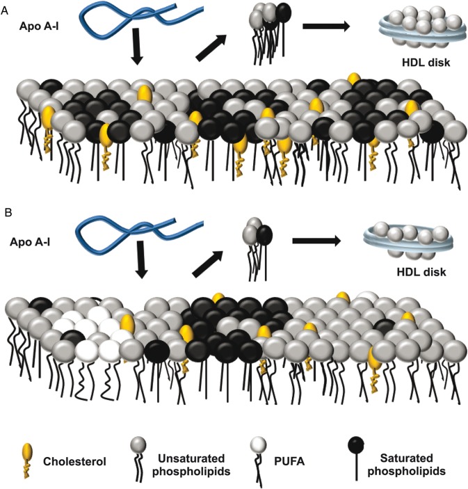
Figure 5.
Schematic representation for the interaction of apoA-I with FADS cells membrane (A) Wild-type PM was represented as domains (rafts) enriched in saturated phospholipids (dark circles with straight legs), segregated from areas enriched in unsaturated phospholipids (gray circles with flexible legs) with Chol molecules (orange oval head with one leg) distributed according to their chemical affinities. (B) The increased PUFA (white circles with flexible legs) content in FADS cells enhanced the segregation of Chol into raft domains. This effect together with a lower content of Chol and SM observed in FADS cells would decrease the surface defect area among different domains. Lipid-free apoA-I (blue ribbon) would interact with both types of PM (black arrows in A and B) solubilizing lipids preferentially from the areas with high surface defects giving rise to disk-shaped particles (blue disks). Thus the lower surface areas in the FADS cells would decrease the efficiency of apoA-I to remove lipids.

