
Figure 2. Heat shock results in HSP70 upregulation in supporting cells.
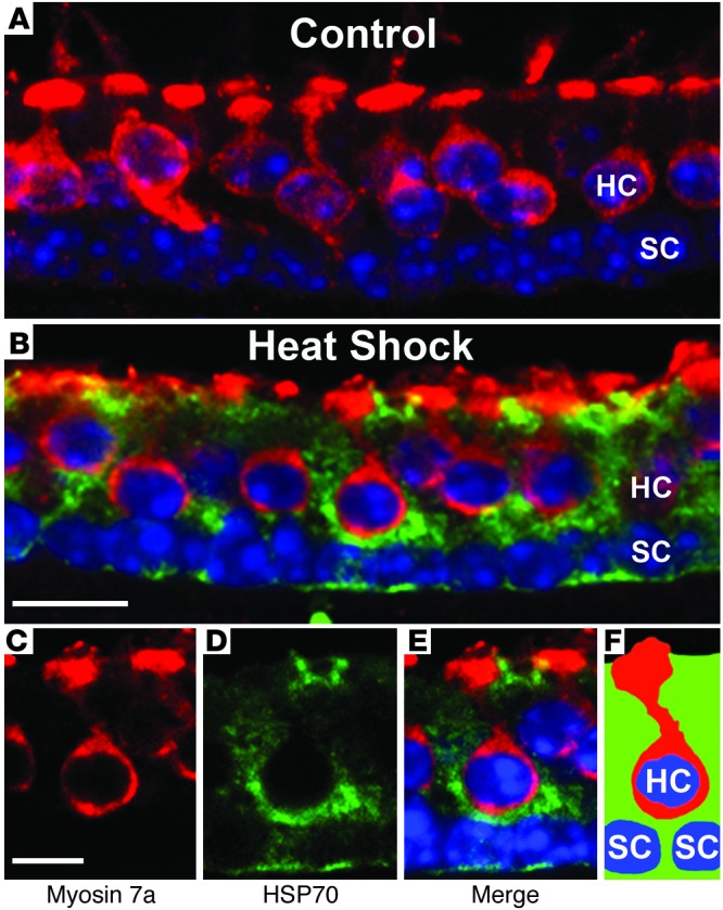
Control and heat-shocked (43°C for 30 minutes) utricles were allowed to recover from heat shock for 6 hours and were sectioned (see Methods). Sections were labeled using antibodies against myosin 7a (red, hair cell marker) and HSP70 (green). Nuclei were labeled with Hoechst dye (blue). Confocal microscopy was performed using identical settings for laser power, gain, offset, and zoom. Shown are a control utricle (A) and a heat-shocked utricle (B–E). Control utricles (A) show very little HSP70 immunoreactivity. Heat-shocked utricles (B–E) show robust upregulation of HSP70 in supporting cells. C–E show a higher-magnification image of a heat-shocked utricle. (C) Myosin 7a labeling of a single hair cell. (D) HSP70 immunoreactivity is present in supporting cells but not in the hair cell. (E) Merged image. (F) Schematic showing locations of the hair cell (HC) and supporting cells (SC) in C–E. Scale bars: 10 μm (A and B); 5 μm (C–E). Images are representative of 3 experiments for a total of 10 to 12 utricles per condition.