Figure 3.

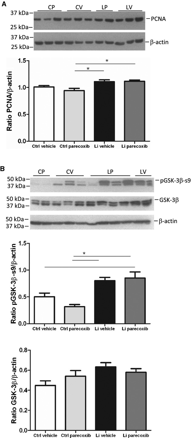
Effect of lithium treatment with and without a COX‐2 inhibitor on cell proliferation and glycogen synthase kinase‐3β phosphorylation. (A) Western blotting (upper panel) of kidney cortex tissue homogenates for proliferating cell nuclear antigen (PCNA), a marker of mitosis, and densitometric evaluation of gels (diagram). Values were normalized to β‐actin protein abundance. Lithium increased PCNA protein abundance with no effect of parecoxib treatment. Data were analyzed with two‐way ANOVA followed by Bonferroni's multiple comparisons test. The columns show mean values ±SEM, *P < 0.05; n = 5 in all groups. (B) Western blotting of kidney cortex tissue homogenates for phospho‐serine 9 (pGSK‐3β‐s9) and total GSK‐3β (upper panel) and densitometric evaluation of gels (diagrams). For samples showing double bands, the upper band was chosen for quantification according to the predicted size of the protein. Lithium treatment significantly increased the abundance of pGSK‐3β‐s9. No effect of parecoxib treatment was seen. The abundance of total GSK‐3β was not significantly altered between any of the four experimental groups. Data were analyzed with two‐way ANOVA followed by Bonferroni's multiple comparisons test. Data are presented as means ± SEM, *P < 0.05; n = 5 in all each group.
