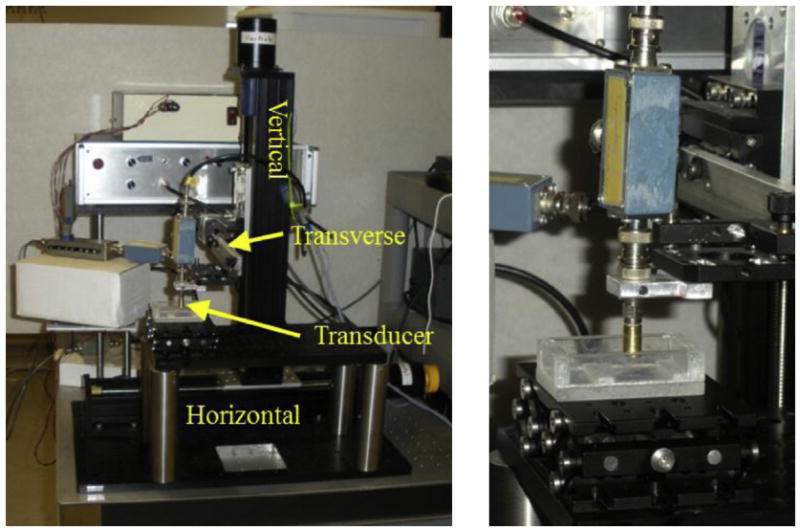
Fig. 1.

Photograph of elasticity microscope equipment. There are three imaging axes: horizontal, vertical and transverse. The deformation axis and slit pushing plate are not shown. On the right is a close-up of the high-frequency ultrasonic transducer (brass-colored cylinder).
