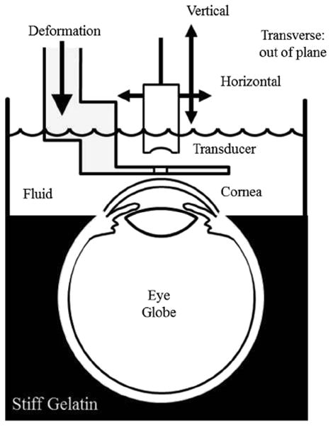
Fig. 2.

Diagram of experimental setup. The eye globe is held in place by gelatin up to the corneal/scleral junction. The rest of the breaker is filled with coupling fluid consisting of water and edema-inhibiting chemicals. Attached to a three-axis scanning system, the single-element transducer images through a slit in the deformation plate that is attached to a fourth motion axis. Machined from aluminum, the deformation plate is about 1 mm thick near the slit.
