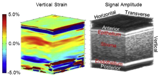
Fig. 3.

Three-dimensional image of vertical strain (left) with a conventional B-mode volume image (right) to highlight corneal anatomy. On the B-mode image, the top bright horizontal/transverse band is the fluid/epithelial boundary. The next bright horizontal/transverse band is the epithelial/stromal boundary. On the bottom the bright band is the endothelial/aqueous boundary. Above the epithelium and below the endothelium is noise from anechoic fluids for both B-mode and strain. Vertical strain in the epithelium is mostly green, indicating little deformation. Along the vertical direction (from anterior to posterior), strain in the stroma gradually transitions from positive to negative.
