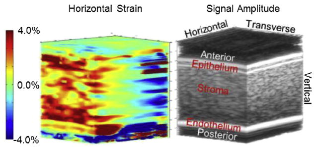
Fig. 5.

Three-dimensional image of horizontal strain (left) with a conventional B-mode volume image (right) to highlight corneal anatomy. The B-mode image is the same as shown in Figure 3. Note that the strain range is slightly smaller than for vertical strain. Positive strain is occurring in half the volume and negative strain in the other half.
