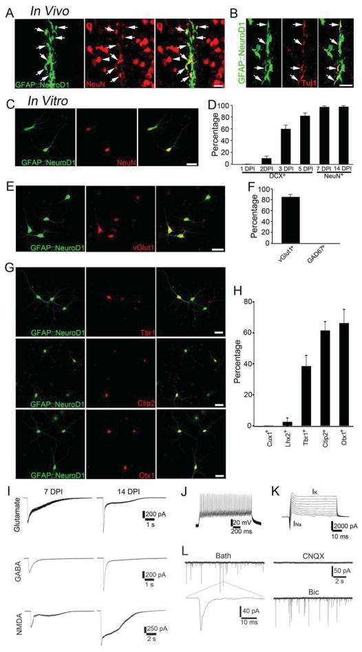
Figure 2. NeuroD1 converts astrocytes into glutamatergic neurons.
(A–B) In vivo injection of GFAP promoter-driven NeuroD1-IRES-GFP (green) retrovirus revealed astrocyte-converted neurons immunopositive for NeuN (A) and Tuj1 (B). (C) Cultured mouse cortical astrocytes were converted into NeuN-positive neurons. (D) Time course of GFAP::NeuroD1 conversion efficiency after infecting cultured mouse astrocytes. (E–F) Astrocyte-converted neurons were positive for VGluT1 (E) but negative for GAD67 (F). (G–H) Immunostaining with cortical layer neuronal markers showed deep layer neuronal properties (Ctip2 and Otx1) after NeuroD1-induced conversion. Scale bars: 20 μm for (A–C) and (E), and 40 μm for (G). (I) Mouse astrocyte-converted neurons showed large glutamate, GABA, and NMDA receptor currents within 2 weeks after NeuroD1 infection. Average GABA current, 7 DPI, 405 ± 97 pA, n=8; 14 DPI, 861 ± 55 pA, n=13. Average glutamate current, 7 DPI, 517 ± 145 pA, n=7; 14 DPI, 1060 ± 159 pA, n=9. Average NMDA current, 7 DPI, 676 ± 118 pA, n=7; 14 DPI, 1315 ± 95, n=7. (J–K) Mouse astrocyte-converted neurons showed repetitive action potentials (J) and large INa and IK (K). (L) Spontaneous synaptic events recorded from mouse astrocyte-converted neurons. All events were blocked by CNQX but not Bic, suggesting that they were glutamatergic events. Also see Suppl. Fig. 4.

