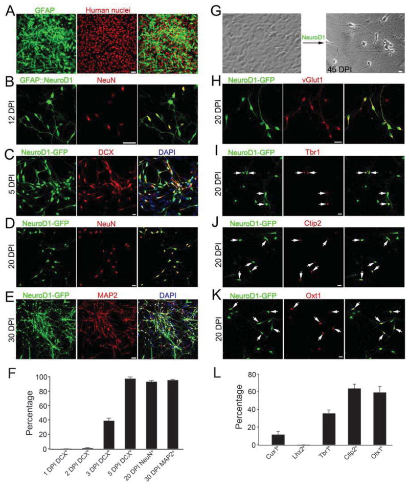
Figure 5. Conversion of cultured human astrocytes into functional neurons.
(A) The majority of cultured human astrocytes were labeled by GFAP (green). (B) Infection by GFAP::NeuroD1 retrovirus converted human astrocytes into NeuN-positive neurons. (C–E) NeuroD1-induced conversion of human astrocytes into neurons as shown by a series of neuronal markers DCX (C), NeuN (D), and MAP2 (E). (F) Quantified data showing a significant increase of conversion efficiency during 3 – 5 DPI. (G) Phase contrast images showing NeuroD1-induced morphological change from astrocytes (left) to neurons (right, 45 DPI). (H) Human astrocyte-converted neurons were immunopositive for VGluT1. (I–K) Cortical layer neuronal markers revealed that human astrocyte-converted neurons were immunopositive for Tbr1 (I), Ctip2 (J), and Otx1 (K). (L) Quantitative analysis of human astrocyte-converted neurons labeled by superficial (Cux1 and Lhx2) or deep layer (Ctip2 and Otx1) neuronal markers. Scale bars: 50 μm for (A) and (E); 20 μm for panels (C–D) and (G–K); 40 μm for panel (B). See also Suppl. Fig. 5–6.

