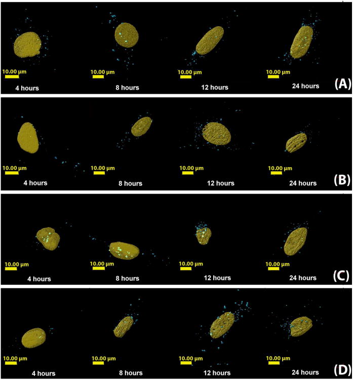
Figure 3.
Three-dimensional reconstructed images of cells transfected with polyplexes formulated with Cy5-pDNA and (A) jetPEI™, (B) Glycofect™, (C) Tr455 and (D) Tr477. With each polymer type, the cells were fixed after 4, 8, 12, and 24 hours. The nucleus is shown in gold and the polyplexes (detected by imaging Cy5-labeled plasmid DNA) are shown in blue. Note: In the time series A-D, the nucleus was set as transparent to view colocalizing polyplexes. The polyplexes colocalizing with the nucleus appear ‘light blue/white’ in color. The controls were cells only, FITC only, DAPI only, Rab 5 primary antibody only, Rab 7 primary antibody only and secondary antibody only. The parameters measured were: (1) volume (μm3) of polyplexes and was determined by the volume of a voxel multiplied by the number of voxels in each physical volume; (2) distance of the polyplex/pDNA complexes from the surface of the nucleus (μm).

