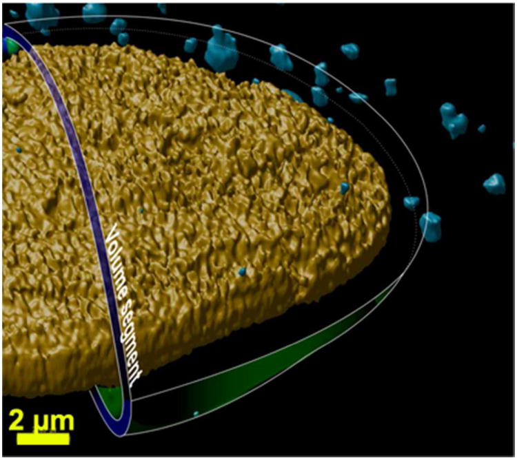
Figure 4.
Proposed concept of a concentric-nuclear zone traversed by the polyplexes in the cell. The image was generated by compiling the two-dimensional confocal microscopy images and three dimensionally rendering the data allowing visualization of the nucleus (yellow) and polyplexes (blue). This image also shows an illustration of an intracellular three-dimensional concentric-nuclear zone (in blue-green) in the perinuclear space to help visualize how the “polyplex distance from the nucleus” was determined.

