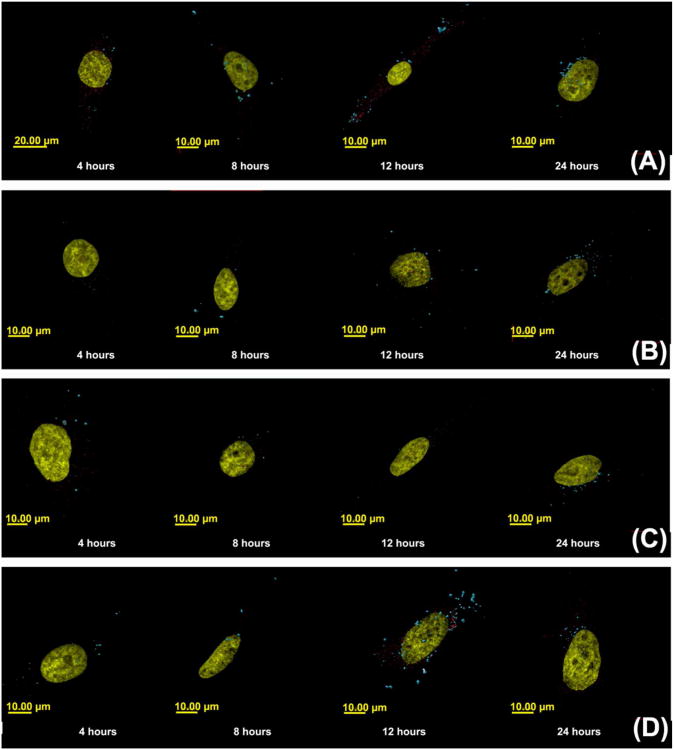
Figure 9.
Colocalization of polyplexes with Rab-7 proteins, a marker for late endosomes. Images A-D show confocal images of HeLa cells monitoring colocalization of polyplexes with Rab 7 proteins for late endosomes at 4, 8, 12 and 24 hour timepoints for polyplexes formed with Cy5-pDNA and (A) jetPEI™, (B) Glycofect™, (C) Tr455 and (D) Tr477. Note: The nuclei are pseudo colored in yellow, the polyplexes are pseudo colored in blue, and Rab 7 is pseudo colored in red. The z-axis is directed into the plane of image which shown in the x-y plane. The parameter measured was 3D colocalization of the polyplex/pDNA with Rab 7 (late endosome marker) in arbitrary units), which was determined by the total of overlapping volumes, rendered from the fluorescence, between the two channels.

