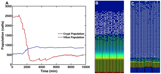
Figure 9. Colonic metaplasia in the ileal pouch.
Panel A displays average crypt and villus gut epithelial cell (GEC) populations after exposure to sustained low-level toll-like receptor (TLR4) stimulation and signaling (an abstraction of fecal stasis). This low-level up-regulation of inflammation communicates via our hypothesized Phosphotase and tensin homolog (PTEN) mechanisms, leading to increased apoptosis, shortening the villus, as well as an inhibition of the Sonic Hedgehog homolog (Hh) pathway, which increases the size of the proliferative compartment (i.e. crypt). Panel B displays a screenshot from SEGMEnT when simulating conditions leading to colonic metaplasia. Crypt hyperplasia and villus atrophy are clearly evident (compare with normal homeostatic condition in Panel C, and as seen in Figure 4C), along with a shift in the villus to crypt height ratio that matches the alterations seen in colonic metaplasia as reported in Ref [27].

