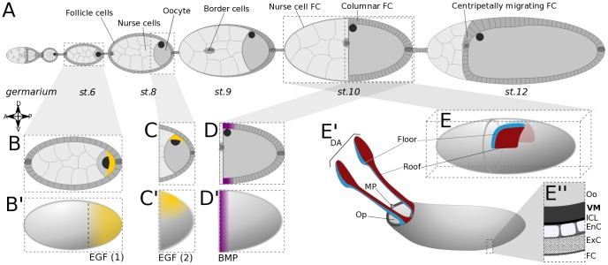
Figure 1. Overview of oogenesis in Drosophila melanogaster.
(A) Schematic of an ovariole. Egg chambers, displayed at progressively later stages from anterior (left) to posterior (right), are formed in the germarium, and consist of three main cell types: nurse cells and the oocyte, both germ line, enveloped by a layer of somatic follicle cells (FC). After stage 9, the FCs have remodeled to form a columnar epithelium over the oocyte, and a squamous epithelium over the nurse cells. (B–B′) At early stages, ligand Gurken (Grk; in yellow) co-localizes with the oocyte nucleus to the posterior pole of the oocyte. It signals to EGFR in the overlying FC, activating the EGF pathway in a posterior-anterior gradient. (C–C′) After oocyte repolarization, Grk and the oocyte nucleus are located at the dorsal-anterior cortex of the oocyte. The EGF pathway is locally activated in overlying FC. (D–D′) Dpp ligand produced in the anterior FC establishes a steep anterior-posterior gradient of BMP signaling activity in the columnar FC. (E–E″) The appendage primordia are defined at stage 10 and consist, on either side of the midline, of two groups of cells, roof and floor. The eggshell deposited between the oocyte (Oo) and the follicle cells (FC) contains the operculum (OP), the micropyle (MP), and two dorsal appendages (DA); and is constituted by the vitelline membrane (VM), the inner chorionic layer (ICL), an endochorion (EnC) and an exochorion (ExC) [31].

