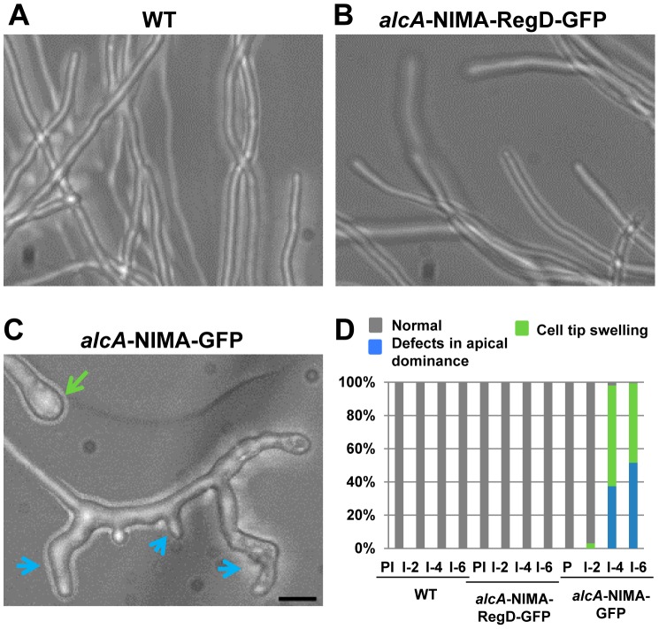
Figure 6. Induction of ectopic NIMA results in defects in tip cell morphology.
(A) Hyphae of wildtype cells or (B) cells carrying alcA driven expression of full length NIMA (alcA::NIMA-GFP, strain: CDS683) or (C) the C-terminal regulatory domain (alcA::NIMA-RegD-GFP, strain: CDS131) were grown under non-inducing conditions and expression of the respective NIMA constructs was induced by the addition of threonine. Representative images of cells after 6 hours of induction are shown. Breakdown of apical dominance and cell tip swelling in NIMA-GFP expressing cells are indicated by blue and green arrows respectively. (D) Quantitation of tip growth defects. PI = Pre-induction, I = alcA induced for 2, 4 or 6 hours as indicated. Bar, 5 μm.

