Fig. 6.
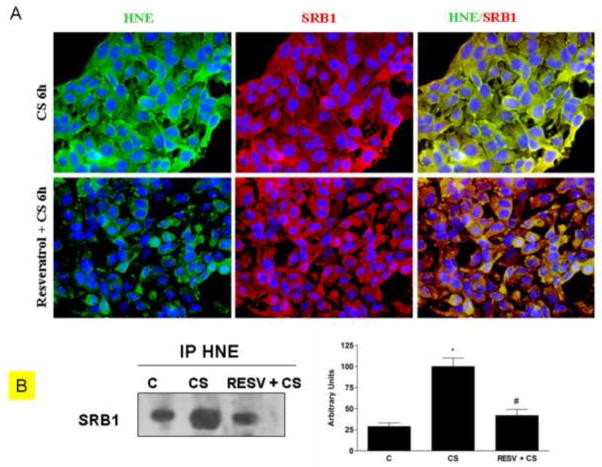
Effect of resveratrol on HNE/SRB1 adducts induced by CS. Immunocytochemistry of HaCaT cells showing localization of HNE-adducts (left column, green color), SR-B1 (central column, red color) and HNE/SR-B1 adducts (right column, yellow color) after Resveratrol pre-treatment and CS exposure(A). Images are merged in the right panel and the yellow color indicates overlap of the staining. These data were confirmed by immunoprecipitation for SR-B1 (B). HaCaT cells were pre-treated with resveratrol and then exposed to CS and cell lysates were immunoprecipitated using anti-HNE. Immunoprecipitated proteins were separated by SDS-PAGE, and then transferred to a nitrocellulose membrane and immunoblotted with anti-SRB1. Western blot shown is representative of five independent experiments. Quantification of the SR-B1 bands is shown on the right of the panel. Data are expressed in arbitrary units (averages of five different experiments ± SEM, * vs. control; # vs. CS; p < 0.05).

