FIGURE 4:
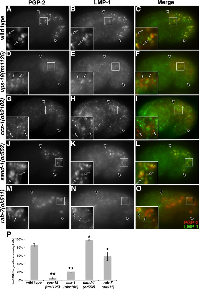
The distribution of PGP-2 and LMP-1 is altered in vps-18, ccz-1, and rab-7 mutants. Antibodies recognizing PGP-2 and LMP-1 colocalized at gut granules in (A–C) wild type and (J–L) sand-1 mutants (black arrows in insets). (D–I) PGP-2–stained organelles in vps-18 and ccz-1 mutants lacked LMP-1 staining (white arrows within insets). (M–O) In rab-7 mutants, LMP-1 staining was weakly present on some (black arrows in insets) but not all (white arrows in insets) anti-PGP-2–marked gut granules. In A–O, 1.5-fold-stage embryos are shown, black arrowheads flank the intestine, and insets are 5 μm wide. (P) For each genotype, at least 25 randomly selected PGP-2–stained intestinal compartments in five different 1.5-fold-stage embryos were scored for the presence of LMP-1 staining. The mean is plotted, and error bars represent the 95% confidence limit. A one-way ANOVA comparing each mutant to wild type was used to calculate p values (*p ≤ 0.05, **p ≤ 0.001).

