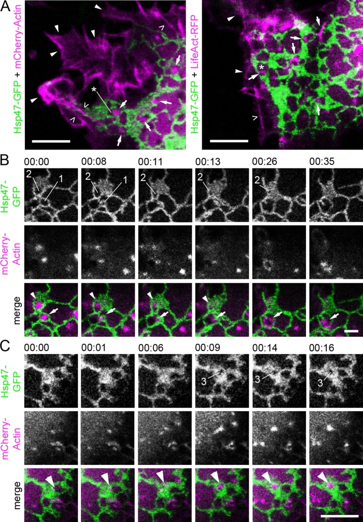
FIGURE 3:
Actin filament arrays localize to polygons defined by the surrounding ER network and have a role in ER sheet movements in Huh-7 cells. (A) Confocal section of live cell coexpressing Hsp47-GFP (green) and mCherry-Actin (magenta) or LifeAct-RFP (magenta). Cortical actin (arrowheads), stress fibers (open arrowheads), and actin filament arrays and foci (arrows) localizing to the polygons (asterisks) are indicated. See also Supplemental Video S3. Time-lapse confocal frames of a cell coexpressing Hsp47-GFP (green) and mCherry-actin (magenta) show that (B) relocation and disappearance and (C) formation of actin arrays and the ER sheet dynamics are interdependent. In B, actin foci (arrow) relocate from polygon (1) to the adjacent polygon at 00:08, leading to subsequent polygon (1) closure at 00:11. Disappearance of actin foci (arrowhead) at 00:13 leads to subsequent polygon (2) closure. See also Supplemental Video S4. In C, arrowhead indicates formation of actin filament array leading to polygon (3) opening within the ER sheet at 00:09. See also Supplemental Figure S3 and Supplemental Video S5. Bars, 5 μm.

