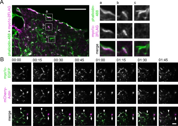
FIGURE 4:
Myo1c localizes and moves in conjunction with actin arrays in Huh-7 cells. (A) Wide-field image of a lamella of fixed cell expressing myo1c-2FLAG, immunolabeled with anti-FLAG antibody (magenta), and stained with phalloidin (green). Boxed areas are shown at higher magnification, portraying myo1c-positive actin filaments at the cell periphery (inset a) and in the cytoplasm (inset b). Occasional actin filaments negative for myo1c were found (inset c). Long actin filaments (open arrowheads) were not positive for myo1c. See also Supplemental Figures S2H and S3. (B) Time-lapse confocal frames (time scale, minutes:seconds) of cell coexpressing myo1c-EGFP (green) and mCherry-actin (magenta) taken from cell lamella show that the actin structures can be both dynamic (actin filament array; arrowhead) and static (actin foci; arrow) and that myo1c moves in conjunction with both actin filaments and foci. See also Supplemental Video S6. Bars, 10 μm (A), 5 μm (B).

