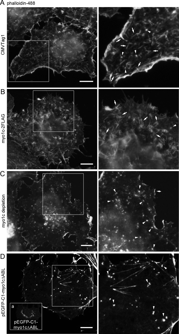
FIGURE 5:
Myo1c regulates actin filament arrays in Huh-7 cells. Wide-field images of fixed cells expressing (A) CMVTag1 (control), (B) myo1c-2FLAG, (D) EGFP-myo1cΔABL, or (C) depleted with myo1c shRNAs were stained with phalloidin. Boxed areas are shown at higher magnification. (D) The EGFP-myo1cΔABL signal is observed as bright spots at the perinuclear area and as diffuse signal throughout the cytoplasm (inset a). See also Supplemental Figures S2E and S4. Bars, 10 μm.

