FIGURE 7:
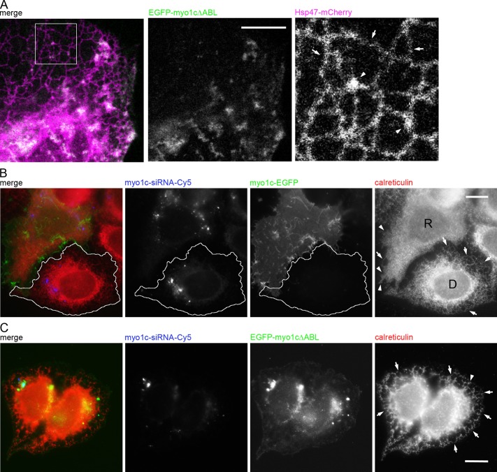
Myo1c actin-binding domain is crucial for proper ER phenotype in Huh-7 cells. (A) Confocal section of a live cell coexpressing Hsp47-mCherry (magenta) and EGFP- myo1cΔABL (green). Tubules (arrows) and the few remaining sheets (arrowheads) are indicated in the magnified image of the boxed area. See also Figure 5D and Supplemental Figure S2I. For rescue experiments, Huh-7 cells were transfected with Cy5-conjugated myo1c siRNA (blue) and, on the next day, siRNA-resistant construct (B) myo1c-EGFP (green) or (C) EGFP-myo1cΔABL (green) and immunolabeled for calreticulin (red). In B, tubules (arrows) and sheets/interlinked ER sheet mass (arrowheads) are indicated in the wide-field images of myo1c-depleted cell (outlined cell; D) and rescued cell (R). In C, the tubules (arrows) of nonrescued cells expressing EGFP-myo1cΔABL are indicated. See Figure 6A for comparison. Bars, 10 μm.

