FIGURE 8:
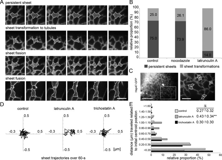
Depolymerization of ER-associated actin filaments increases sheet transformations and sheet fluctuations in Huh-7 cells. (A) Wide-field time-lapse images of representative sheet (outlined) dynamics of cells expressing Hsp47-GFP. (B) Relative proportions (%) of persistent sheets vs. transforming sheets over the 60-s observation time (1 frame/s) in control and nocodazole- and latrunculin A–treated cells. (C) Still frame of a wide-field video subjected to center-of-mass analysis. The boxed area of a cell expressing Hsp47-GFP is shown at higher magnification on right, where the manually traced sheets and their calculated centroid (asterisks) are indicated. (D) Representative trajectories of sheet centroid movements over 60 s, plotted in a 1-μm2 area, of control and latrunculin A– and trichostatin A–treated cells and (E) the subgrouped centroid distances (μm) to the initial centroid position plotted against occurrence (%) and average distance ( , μm ± SD) to the initial centroid position. The lateral movement increased significantly (**p < 0.05) with latrunculin A but not trichostatin A treatment compared with controls. Bars, 2.5 μm (A and magnified image in C), 10 μm (otherwise in C).

