
FIGURE 3:
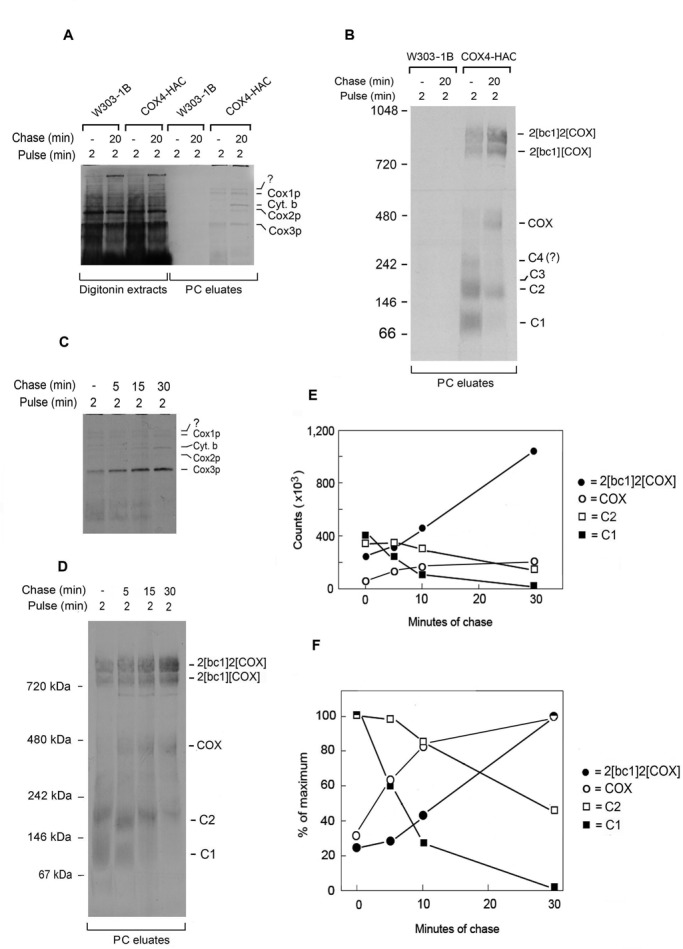
Pulse-chase analysis of Cox4p-HAC incorporation into COX. (A) W303-1B (WT) and W303/COX4-HAC (COX4-HAC) expressing tagged Cox4p were grown to early stationary phase in YPGal with additional 2 h of growth in YPGal containing 2 mg/ml chloramphenicol. Mitochondria (0.5 mg of protein) were labeled for 2 min as in Figure 1A. Half of the translation mixture was extracted with digitonin immediately after addition of puromycin and cold amino acids (0-min chase). The other half was allowed to incubate for an additional 20 min (20-min chase) before extraction with digitonin. The extracts were purified on protein C antibody beads as in Figure 1A, and samples of the extracts (6% of total) and eluates (PC eluates) from the antibody beads (15% of total) were separated by SDS–PAGE on a 17% polyacrylamide gel and radiolabeled proteins visualized as in Figure 1A. The radiolabeled bands in the eluates are identified in the margin. (B) The remainder of the eluates (80% of total) from A was separated by BN-PAGE on a 4–13% polyacrylamide gel and radiolabeled proteins visualized. (C) Mitochondria of W303/COX4-HAC were pulse labeled for 2 min and chased for the indicated times as in A before extraction with digitonin. Digitonin extracts were purified on protein C antibody beads and separated by SDS–PAGE on a 17% polyacrylamide gel. (D) Samples purified on antibody beads were separated by BN-PAGE on 4–13% polyacrylamide gel and radiolabeled proteins visualized. (E, F). The radiolabeled bands in D were quantified in a phosphorimager.