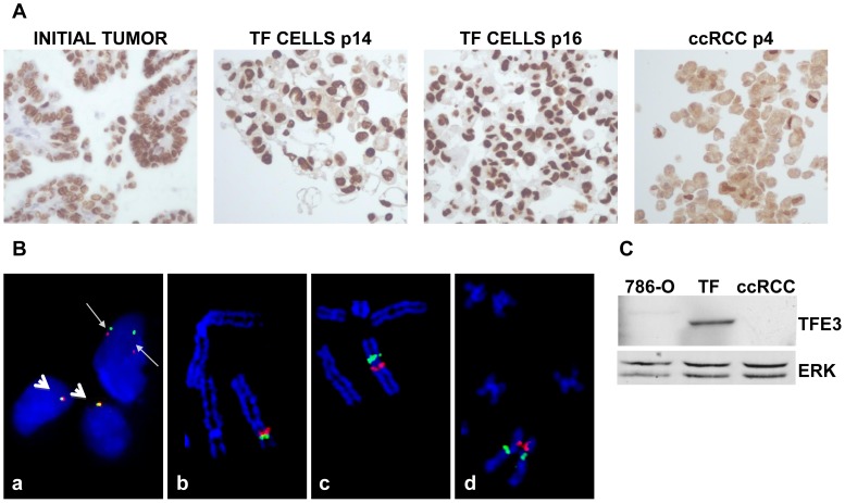
Figure 1. Characterization of TFE3 expression and TFE3 rearrangement in the initial tumor and in TF cells.
A) Immunohistochemical staining for TFE3 of the initial tumor. Labeling with anti-TFE3 antibodies was also performed on cells from passages 14 and 16 (P14 and P16 TFE3 cells) embedded in paraffin. TFE3 labeling was also performed on ccRCC cells cultured under the same conditions as TF cells. ccRCC cells served as a negative control. Note the cytoplasmic background instead of only nuclear labeling. B) Image a: An uncultured cell suspension from the renal cell tumor hybridized with a dual-color break-apart FISH probe framing TFE3. A rearrangement of TFE3 in the upper nucleus (tumor cell) is observed with BAC probes CTD-2534B7 (red signal; 3′ side of TFE3) and CTD-3009K20 (green signal; 5′ side of TFE3). The red and green signals are clearly separated as a result of the pericentric inversion of chromosome X (thin arrows). Two rearranged signals are observed because the abnormal chromosome X is duplicated in tumor cells. In contrast, red and green signals are closely juxtaposed in the two normal nuclei that contain one X chromosome, respectively (thick arrows). Image b: A normal partial metaphase cell hybridized with a dual color break-apart BAC FISH probe framing TFE3. BAC probes CTD-2534B7 (red signal; 3′ side of TFE3) and CTD-3009K20 (green signal; 5′ side of TFE3) are closely juxtaposed at Xp11.23 on the short arm of the X chromosome. Image c: A partial abnormal tumor metaphase cell (cell line, passage 9) hybridized with a dual color break-apart FISH probe framing TFE3. As a result of the X chromosome pericentric inversion, BAC probe CTD-3009K20 (green signal; 5′ side of TFE3) is translocated from its normal location at Xp11.23 to NONO locus at Xq13.1 on the long arm of the X chromosome. BAC probes CTD-2534B7 (red signal; 3′ side of TFE3) remains at the original TFE3 locus at Xp11.23. Image d: A partial abnormal tumor metaphase cell (cell line, passage 9) hybridized with a dual-color break-apart FISH probe framing NONO. As a result of the X chromosome pericentric inversion, BAC probe RP11-753F2 (green signal; 5′ side of NONO) is translocated from its normal location at Xq13.1 to TFE3 locus at Xp11.23 on the short arm of the X chromosome. BAC probes RP11-624G23 (red signal; 3′ side of NONO) remains at the original NONO locus at Xq13.1. C) Western blot analysis of the presence of TFE3 in cells from the “TFE3” tumor, in ccRCC 786-O cells and ccRCC cells obtained from an independent tumor. 786-O and ccRCC cells served as negative controls. ERK served as a loading control.

