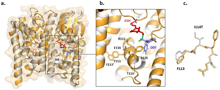
Figure 2. Crystal structure of GSTE2 ZAN/U variant.
a. Superposition of the crystal structure of ZAN/U determined in this study (orange) and the Kimusu 1B variant (grey; PDB entry IMI). A high degree of local and overall structural agreement is clearly noticeable. The location of the docked DDT is based on the computational prediction of Wang et al.[15]. Some manual adjustments were made to relieve steric clashes and to better superimpose the DDT on the position of the hexyl group of bound S-hexylglutathione. b. Close-up detail of the ZAN/U active site. c. Superposition of structure of ZAN/U and Kimusu 1B variant local to position 114 (colour code as in a. A superimposition of ZAN/U from An. gambiae with the GSTE2 from An. funestus is provided in Figure S3).

