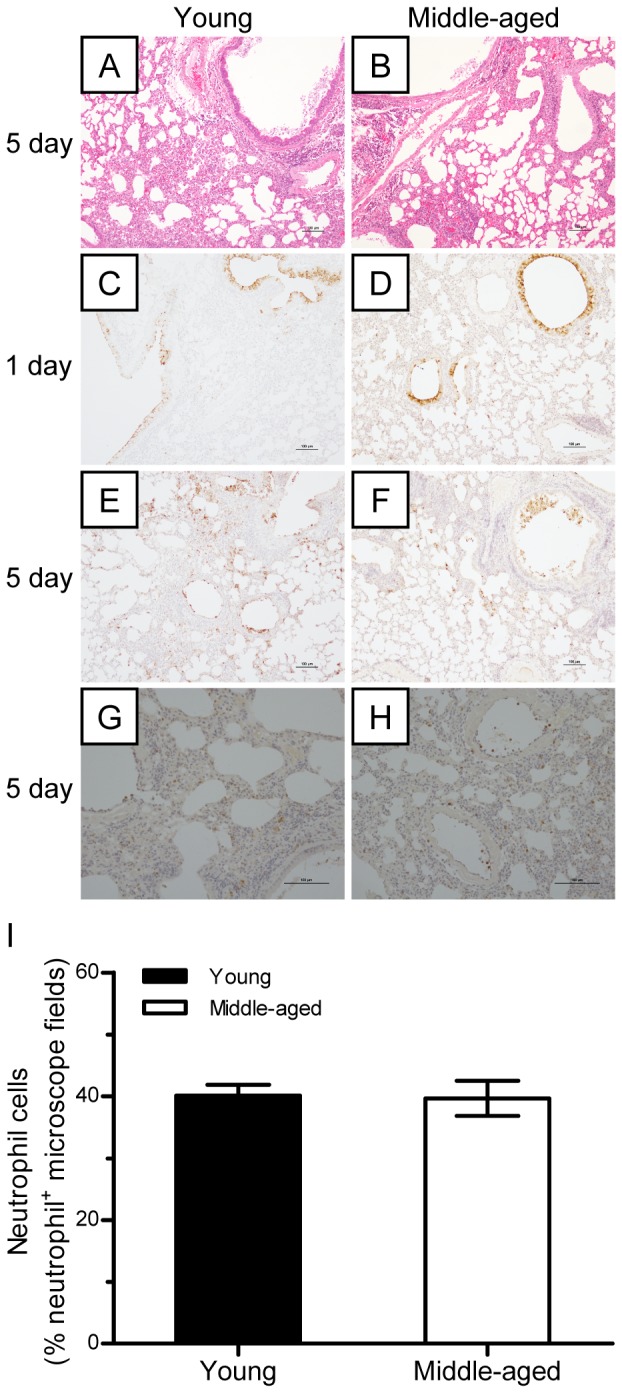
Figure 7. Lung lesion and virus replication in young and middle-aged mice.

(A–B) Young and middle-aged BALB/c mice were challenged intranasally with 106 TCID50 virus and euthanized at days 1 and 5 postviral infection. (A–B) Histological analysis of the lung tissues of mice on days 1 and 5 postviral infection. (C–F) Virus antigen distribution in the lungs on days 1 and 5 postviral infection (n = 5). (G–I) Neutrophil infiltration in the parenchyma of the lungs on days 1 and 5 postviral infection (n = 5).
