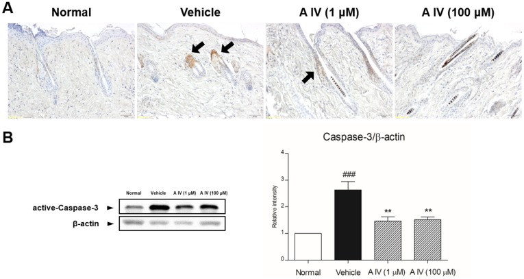
Figure 3. Expression of caspase-3-positive cells by immunohistochemistry (A) and activation of caspase-3 by western blot (B).
The magnification is ×200. Arrows point to caspase-3-positive cells. Results are presented as mean ±S.D. ## indicates the mean differs significantly between normal group and depilated group (p<0.01). * indicates that the mean differs significantly between Astragaloside IV-treated group and depilated group (p<0.05).

