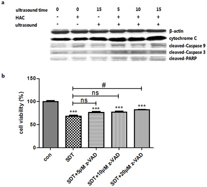
Figure 8. Protein levels changes were analyzed by western blot.
(a) Protein levels of cytochrome C, cleaved-Caspase-9, cleaved-Caspase-3 and cleaved-PARP in the control and 5.0 μg/mL HAC alone, ultrasound alone, SDT (5.0 μg/mL HAC plus 5, 10 and 15 minutes ultrasound exposure) groups. β-actin was used as a loading control. (b) Effects of Caspase inhibitor z-VAD on cell viability of THP-1 macrophages. The cells were detected by CCK-8 assay after 6 h incubation following SDT with or without z-VAD pre-treatment. ***p<0.001 vs. control. #p< 0.05 vs. SDT.

