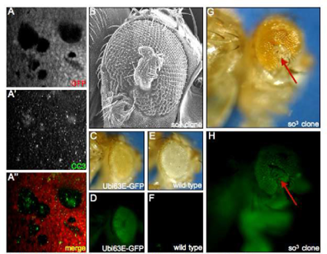
Figure 4. Fate of so and eya Mutant Tissue in the Developing and Adult Retina.

(A) Immunofluorescence images of a so3 mutant clone. Immunostained cleaved Caspase-3 is labeled green and GFP is labeled red. (B). Scanning electron micrograph of a cuticular outgrowth emanating from the compound eye. (C,D) Light and fluorescent images of an adult fly harboring a GFP reporter under the control of the Ubi63E promoter. Note that GFP is expressed in both the retina and the surrounding cuticle. (E,F) Light and fluorescent images of a fly lacking the Ubi63E-GFP transgene. Note that GFP expression is not observed. (G, H) Light and fluorescent images of an adult fly harboring so3 mutant clones. Red arrows point to cuticular outgrowth from the eye. Note the presence of GFP in the cuticular outgrowth. Genotypes are listed at the bottom right of each panel. Anterior is to the right.
