
Figure 7. Models for So-Eya-Dac Regulation.
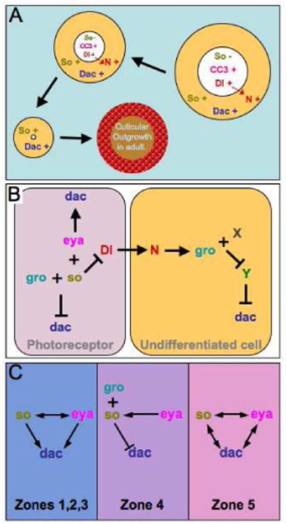
(A). Schematic of the events observed in so or eya clones located in zone 4 of the developing eye disc. Note that the mutant tissue in the disc undergoes programmed cell death and is eliminated by the adult stage and is denoted by the shrinking size of the clone. The cuticular outgrowth in the adult is derived from the dac positive undifferentiated cells that surround the mutant clones in the eye disc. (B) A schematic depicting a potential mechanism for the repression of dac in photoreceptor neurons and undifferentiated cells in zone 4 of the retina. (C) A series of models describing the genetic interactions that we observe between so, eya and dac in the developing retina. Note that the relationships change depending upon the spatial orientation within the eye field.