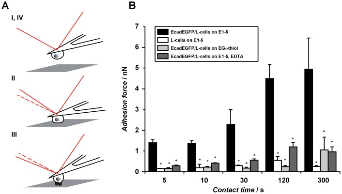
Figure 4. Single-cell force spectroscopy.
(A) Schematic depiction of an SCFS experiment. A single probe cell is attached to an AFM cantilever (I) and brought in contact with the substrate (II). During cell retraction (III) a force curve is recorded from which the cell adhesion force can be determined. (B) Adhesion forces (mean ± SEM) measured at different contact times. EcadEGFP/L-cells on E1-5 (black), on EG4-thiol (light grey), on E1-5 in the presence of 10 mM EDTA (dark grey) and untransfected L-cells on E1-5 (white) were tested. The significance was determined using the Mann-Whitney test (* p< 0.05). At least 10 cells were tested per condition.

