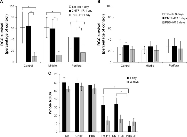
Figure 7.
Statistical analysis of RGC survival following Tet application in vivo.
Notes: (A) Percent survival of RGCs in PBS intravitreal-injected (PBS), 10 μM Tet intravitreal-injected (Tet), 12.5 ng/mL CNTF intravitreal-injected (CNTF), PBS intravitreal-injected plus ischemia (PBS–I/R), and 10 μM Tet intravitreal-injected plus ischemia (Tet–I/R) as well as 12.5 ng/mL CNTF intravitreal-injected plus ischemia (CNTF–I/R) eyes were determined by DAPI stain. Intravitreal injection of Tet 1 day before retinal ischemic reperfusion significantly improved ganglion cell survival one day after ischemia in the central, middle, and peripheral retinal regions (P<0.05). Under the Tet and CNTF conditions (B), there were no significant differences in RGC survival 3 days after ischemia in any of the retinal regions (P>0.05). (A and B) Results are expressed as a percent of the PBS intravitreous injection control. (C) The total RGC numbers in PBS, Tet, CNTF, PBS–I/R, Tet–I/R, and CNTF–I/R eyes 1 day and 3 days after ischemia. (C) The numbers of RGCs in the PBS–I/R, Tet–I/R, and CNTF–I/R retinas 1 day and 3 days after ischemia were much lower than the corresponding numbers in the PBS, Tet, and CNTF groups, respectively (P<0.05). A 2 μL volume of Tet, CNTF, or PBS was intravitreally injected 24 hours prior to ischemia. Results are expressed as means ± SD. *P<0.05 as compared to control, one-way ANOVA. #P<0.05, one-way ANOVA (n≥3).
Abbreviations: ANOVA, analysis of variance; CNTF, ciliary neurotrophic factor; I/R, ischemia/reperfusion; PBS, phosphate-buffered saline; RGC, retinal ganglion cell; SD, standard deviation; Tet, tetrandrine.

