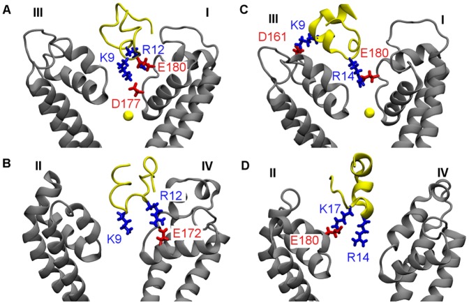
Figure 4. Structures of PIIIA in complex with NaV1.4 predicted from MD simulations biased with distance restraints.
In two binding modes of PIIIA-NaV1.4, Lys9 (A and B) and Arg14 (C and D) of the toxin protrudes into the filter of the channel, respectively. In (A) and (C), the Na+ ion inside the filter is shown as a yellow sphere. Toxin backbone is in yellow and channel backbone in grey.

