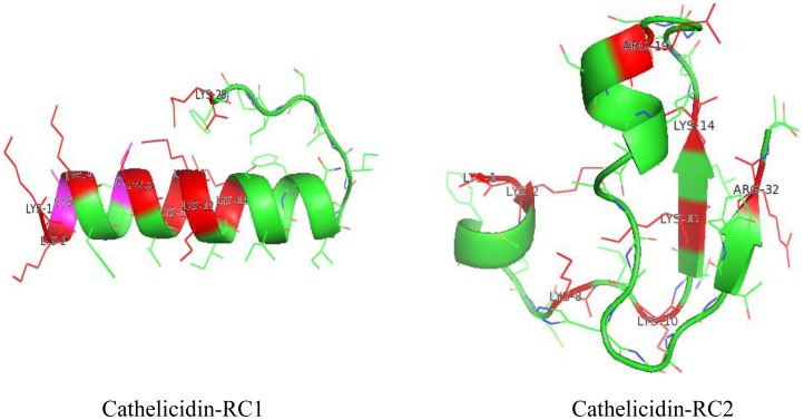
Figure 4. Secondary structure modeling of cathelicidin-RCs.
The models of cathelicidin-RCs were produced by Mod6v2 version of MODELLER. Visualization of the structures were accomplished by Pymol and represented in the form of ribbons. The homology modeled structures were displayed in green. Residues of Lysines and Arginines were labeled in red and Cysteines were labeled in purple in shortened forms.

