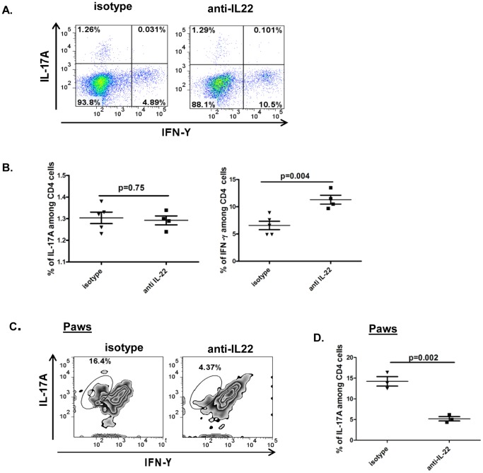
Figure 4. Neutralization of IL-22 after onset of arthritis preferentially increases the frequency of Th1 cells in lymphoid organs but not in joints.
Experimental design is similar to figure 2. Draining inguinal lymph node cells and single cell suspensions of the paws from mice receiving anti-IL-22 antibody or isotype control were briefly stimulated ex-vivo with PMA/ionomycin and Brefeldin A for 6 hours followed by fluorescent labeling for surface anti-CD4 antibody and intra-cellular anti-IL-17A and anti-IFN-γ antibody. A: Representative dot plots shows IFN-γ and IL-17A staining on gated CD4 cells from lymph nodes. B: Percentages of CD4+IL-17+ or CD4+IFN-γ+ lymph node cells from the two groups of mice were plotted as a dot plot with each dot representing an individual mouse. C: Single cell suspension of joint cells from mice receiving anti-IL-22 antibody or isotype control were briefly stimulated ex-vivo with PMA/ionomycin and Brefeldin A for 6 hours followed by fluorescent labeling for surface anti-CD4 antibody and intra-cellular anti-IL-17A and anti-IFN-γ antibody. Representative zebra plots show IFN-γ and IL-17A staining on gated CD4 cells from paws. D: Percentages of CD4+IL-17+ cells from the paws of two groups of mice were plotted as a dot plot with each dot representing an individual mouse.

