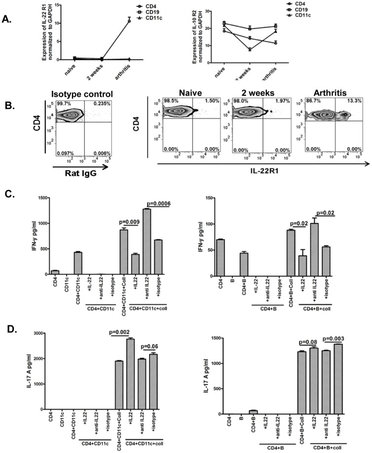
Figure 5. Increased expression of IL-22R1 on CD4 cells from arthritic mice and regulation of IFN-γ responses in T cells by IL-22.
A: Expression of IL-22R1 and IL-10R2 were analyzed by realtime PCR in CD4+, CD19+, and CD11c+ cells enriched from splenocytes from various phases of arthritis. GAPDH was used as internal control. Data represented as fold change over GAPDH expression. Data is representative of 3 independent experiments. B: Splenocytes from various phases of arthritis were stained for CD4, and IL-22R1 and analyzed by flowcytometry. RatIgG was used as isotype control. Data shown is gated on mononuclear cells based on forward and side scatter followed by gating on CD4+ cells. Data is representative of 3 independent experiments. C& D: CD4 T, CD11c, and B cells were enriched from splenocytes of arthritic mice and co-cultured with recombinant IL-22 (100 ng/ml, Insight Genomics, USA), or anti-IL-22 antibody (10 ug/ml) or mouse isotype control (10 ug/ml, Biolegend) and/or collagen for 7 days. Supernatants were analyzed for IFN-γ (Fig. 5C) or IL-17A (Fig. 5D) by ELISA. Data is representative of two independent experiments.

