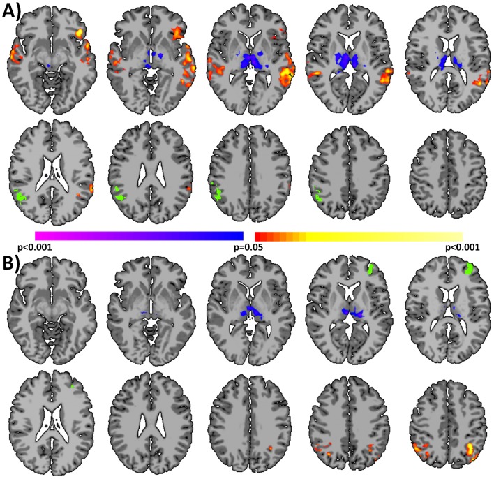
Figure 4. Seed-based connectivity results using seeds detected by coupled-ICD.
A) Several regions of the brain displayed evidence for both increased and decreased connectivity during the anesthesia state as detected by coupled-ICD. A follow-up seed-based analysis on independent data for one of these regions (the right parietal lobe; green region) revealed both significant (p<0.05 corrected) increases and decreases in connectivity to this region, echoing the coupled-ICD results. B) For some regions of the brain, coupled-ICD and conventional approaches showed seemingly conflicting results, with coupled-ICD suggesting decreased connectivity while conventional approaches suggest increased connectivity due to anesthesia. Seed connectivity for one of these regions (the left frontal lobe; green region) revealed both significant (p<0.05 corrected) increased and decreased connectivity, demonstrating that the different approaches may be sensitive to different aspects of changes in connectivity.

