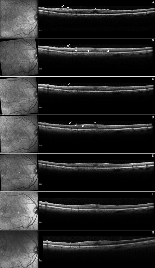
Figure 1.
Infrared picture (left), spectral domain optical coherence tomography (right): Example of a patient imaged with an intense series of spectral domain optical coherence tomography (SD-OCT). A Pre-operative OCT showing the ERM with fibrillation of the nerve fiber layer = NFL (arrow), surface wrinkling (arrowhead) and retinal thickening with foveal evagination (asterix). B Post-operative OCT at day 2, showing still irregulation of the NFL (arrow) and the outer plexiform layer = OPL (arrowheads) . C OCT at week 2 with persisting of NFL fibrillation. D OCT at 1 month showing for the first time normalization of the NFL (arrow) and the reflectivity y of the OPL. Also note the retinal thinning of the fovea with amelioration of the foveal contour (asterix). The OCT images at 3 months (E), 6 months (F) and 9 months (G) do not show any significant signs for retinal change after surgery anymore. The foveal contour was not completely restored in this patient whereas BCVA was already 60 letters at ETDRS charts at month 1.

