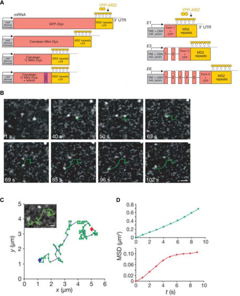
Figure 11. Single particle tracking of mRNPs in mammalian cells.
(A) Schematic of the mRNA constructs with 24xMS2 binding sites used in this study. Nuclear diffusion data for each construct are summarized in Table 2. (B) Deconvolved time-series images of nuclear Cerulean-½ Mini-Dys + intron mRNP diffusing in the nucleoplasm (green tracks). (C) Complete track of the nuclear Cerulean-½ Mini-Dys + intron mRNP. (D) MSD analysis of single nuclear mRNPs displaying Brownian (green) or corralled (red) motions in the nucleoplasm. Corralled motions deviate from linearity at longer lag times. Scale bar represents 1 μm. Reprinted with permission from ref. 225. Copyright 2010 Nature Publishing Group.

