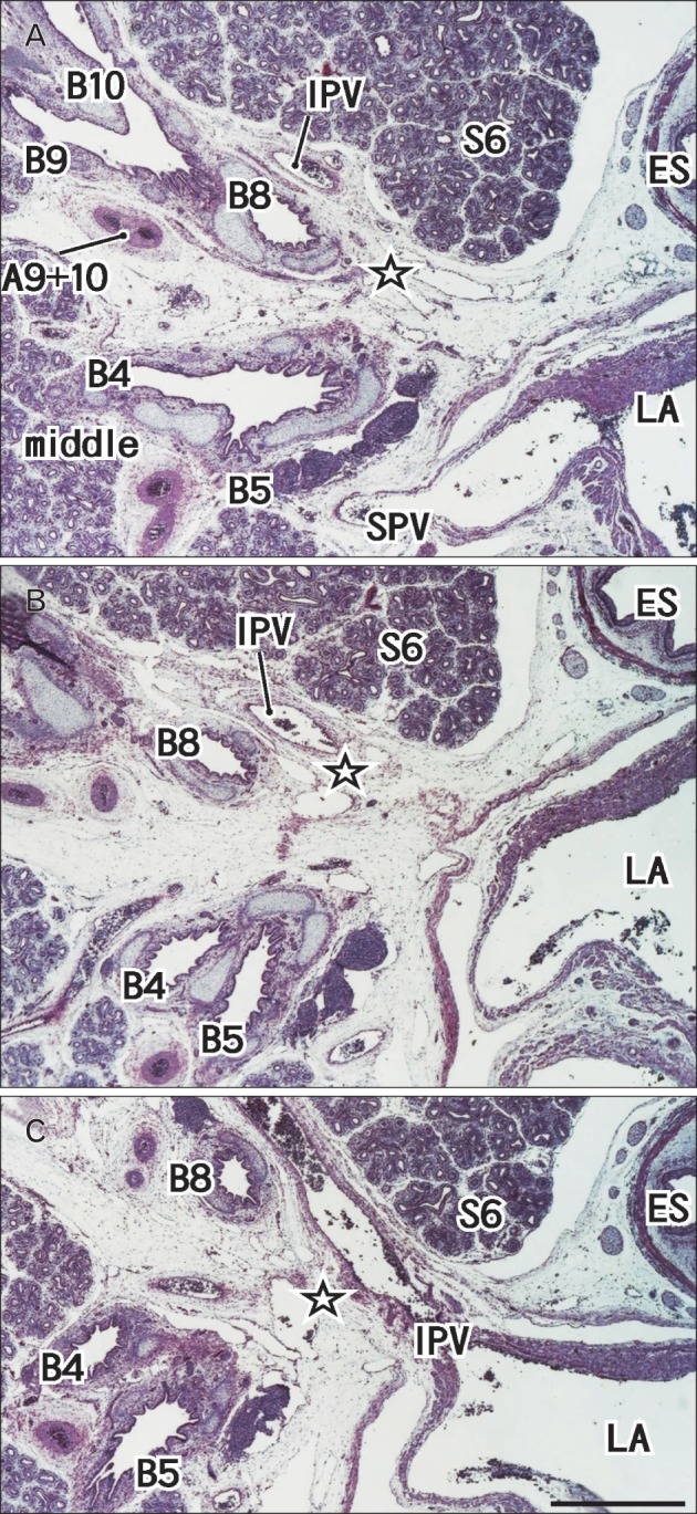
Fig. 3.

A 16-week specimen with segmental bronchus VII. Panel (A) (or C) is the most superior (or inferior) in the figure. Intervals between panels are 0.3 mm (A-B, B-C). Star indicates an area in which the segmental bronchus VII is likely to present in the immediately anterior side of the inferior pulmonary vein (IPV). A9+10, a common trunk artery of the segmental arteries IX and X; B4, B5, B6, B8, B9, and B10, segmental bronchi IV, V, VI, VIII, IX, and X; ES, esophagus; LA, left atrium; S6, segment VI; SPV, superior pulmonary vein. All panels are prepared at the same magnification and include the middle and lower lobes. Scale bar in panel (C)=1 mm (A-C).
