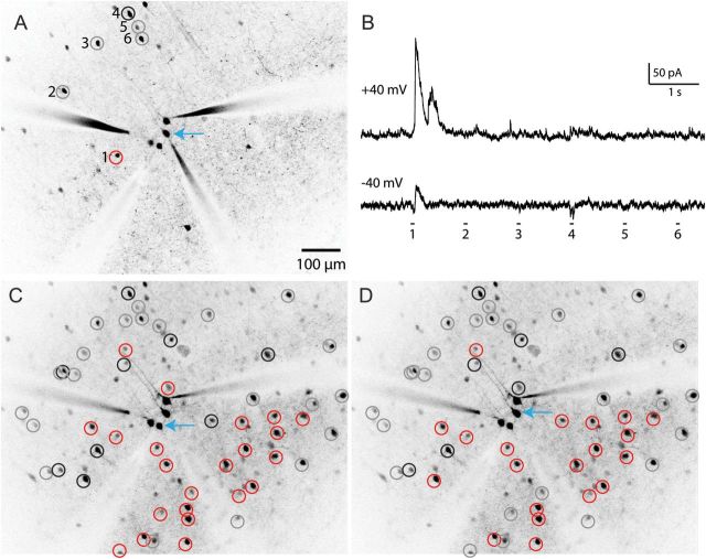
Figure 1.
Mapping inputs from interneurons to pyramidal cells. (A) Two-photon mapping of synaptic connections from 6 PV interneurons (numbered) onto a PC (blue arrow). (B) PV interneurons were photostimulated with 2-photon RuBi-Glutamate uncaging as described previously (Packer and Yuste 2011). Interneurons were sequentially stimulated, while responses were recorded from the postsynaptic PC held at a membrane potential of +40 or −40 mV (upper and lower traces) to distinguish inhibitory from rare but possible excitatory inputs. Note the large, low-latency IPSCs evoked during the stimulation of interneuron 1 (red circle, left), which do not change direction around the glutamate reversal potential, indicating an inhibitory monosynaptic connection from this interneuron. All other interneurons show no response indicating a lack of synaptic connection (gray circles), while interneuron 4, on the other hand, exhibits a weak excitatory response and is labeled as false positive (black circle). (C) Checking all interneurons in this field of view across multiple focal planes yields an inhibitory input map for the PC indicated by the blue arrow. (D) Another input map for a different PC directly adjacent to the PC mapped in C. The other PCs patched in this field did not yield recordings of sufficient quality for mapping.

