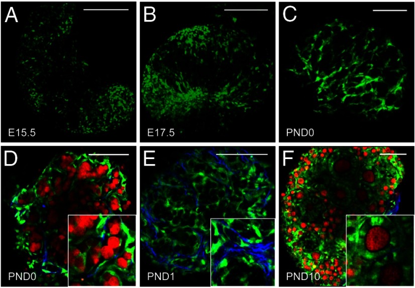
Figure 2.
Notch active cells undergo extensive reorganization during follicle histogenesis. A, Expression of EGFP (green) from the Notch reporter line was first detected in embryonic ovaries at E15.5, although the levels were low. B, At E17.5, Notch active cells can be seen within the ovary, but are not well organized. C, By PND0, Notch active cells are arranged in a cage-like pattern around germ cell syncytia. D, In addition, Notch active cells (green) can be seen encapsulating individual germ cells (red) within syncytia that are labeled with a conditional allele for tdTomato under the direction of Vasa-Cre (63). E, Notch active cells can also be seen along collagen fibrils (blue), detected by second harmonic generation (79). F, EGFP continues to be detected in granulosa cells of primary follicles, although there is variability in the levels detected between neighboring cells. Scale bars, 100 μm.

