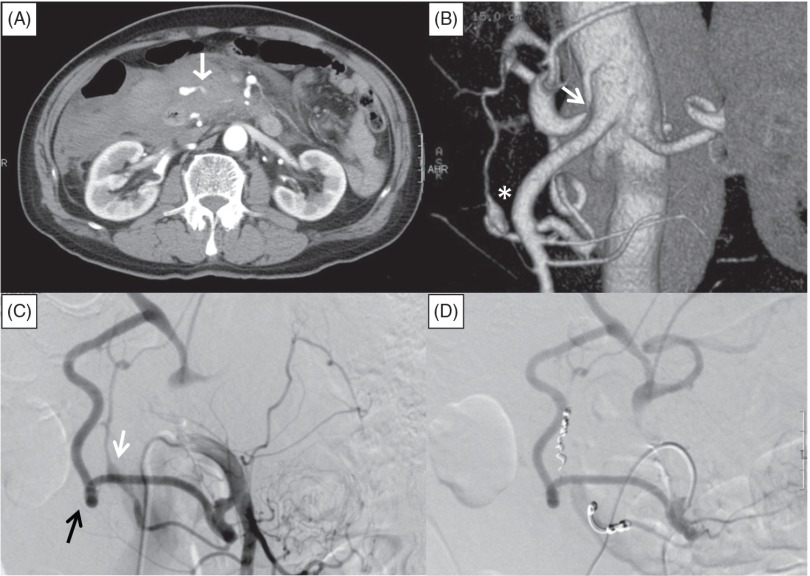
Fig. 1.
Abdominal contrast-enhanced computed tomography (CT) and angiography of the superior mesenteric artery (SMA) in Case 1. (A) Retroperitoneal hematoma with extravasation from an artery (white arrow) adjacent to the dorsal side of the pancreatic head. (B) Three-dimensional CT image showing luminal stenosis of the celiac axis (white arrow) and an aneurysm of the pancreaticoduodenal artery (PDA) (asterisk). (C) Selective catheter angiography of the SMA showing retrograde flow from the SMA to the common hepatic artery via the larger anterior PDA (black arrow) and the smaller posterior PDA with an aneurysm (white arrow). (D) SMA angiography after embolization, showing no flow into the posterior PDA and the aneurysm.

