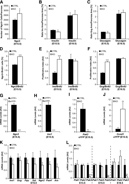
Figure 5.
Increased islet progenitors and β-cell proliferation together with impaired β-cell differentiation in Pak3-deficient embryos. Differentiation (A–C) of endocrine cells was explored at E15.5 and E18.5 in Pak3-deficient (KO) and control mice (CTRL) by immunofluorescence for Ngn3, insulin, and glucagon and quantified. Proliferation (D–F) was assessed and quantified in parallel by evaluating cells in S phase by costaining for BrdU (2-h pulse). The number of Ngn3+ islet progenitor cells increases at E15.5 (A), whereas the number of α- and β-cells is reduced at E15.5, but not at E18.5 (B and C), in Pak3-deficient embryos compared with controls. Ngn3 cells (D) as well as developing α- (F) and β-cells (E) proliferate more in Pak3-deficient embryos at E15.5. G–J: eYFP+ cells were sorted from E15.5 Ngn3eYFP/+ pancreas (littermates) that were either Pak3 KO or heterozygous controls and analyzed by RT-qPCR. Ngn3- and insulin-expressing cells (due to the stability of the eYFP protein) are purified in the eYFP+ population (G and H) and Pak3 is lost as expected in eYFP+ cells from Pak3 KO embryos (I). Transcription of cyclin D1 (Ccnd1) is upregulated in eYFP+ cells from Pak3 KO embryos compared with controls (J). K: RT-qPCR for different endocrine markers in Pak3 KO and control pancreas at E15.5. L: RT-qPCR for Pak1–3 in Pak3 KO and control pancreas from E13.5, E15.5, and E18.5 embryos. For each experiment, three to four control and mutant samples were analyzed. Data are represented as mean ± SD. AU, arbitrary units. B and C: Pancreas area = DAPI area. E and F: AU = number of BrdU/Ins+ or Gcg+ cells/Ins or Gcg area × 10,000. *P < 0.05; **P < 0.01; ***P < 0.001.

