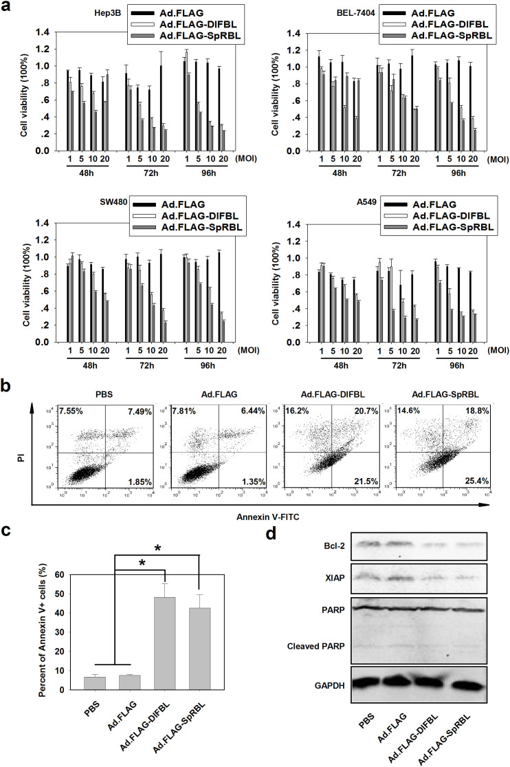
Figure 1. Ad.FLAG-DlFBL and Ad.FLAG-SpRBL induced apoptosis and suppressed cancer cell proliferation.
(a) Hepatocellular carcinoma cell lines Hep3B and BEL-7404, colorectal cancer cell line SW480, and lung cancer cell line A549 were treated with Ad.FLAG, Ad.FLAG-DlFBL, or Ad.FLAG-SpRBL at 1, 5, 10, or 20 MOIs for the time periods indicated. Cell viability was analyzed through MTT assay. Values from at least 6 repeats were calculated as percent of PBS control and presented as mean ± SEM. (b) Hep3B cells treated with Ad.FLAG, Ad.FLAG-DlFBL, or Ad.FLAG-SpRBL at 20 MOI as well as PBS control for 48 h. Cells were then stained with Annexin V-FITC and PI and analyzed under a flow cytometer. (c) The percent of Annexin V-positive cells from 3 repeats were shown as mean ± SEM (*: p < 0.05). (d) Cell lysates were analyzed by Western blot for levels of Bcl-2, XIAP, and PARP. GAPDH served as the loading control. Full length blots were shown in Fig. S1.

