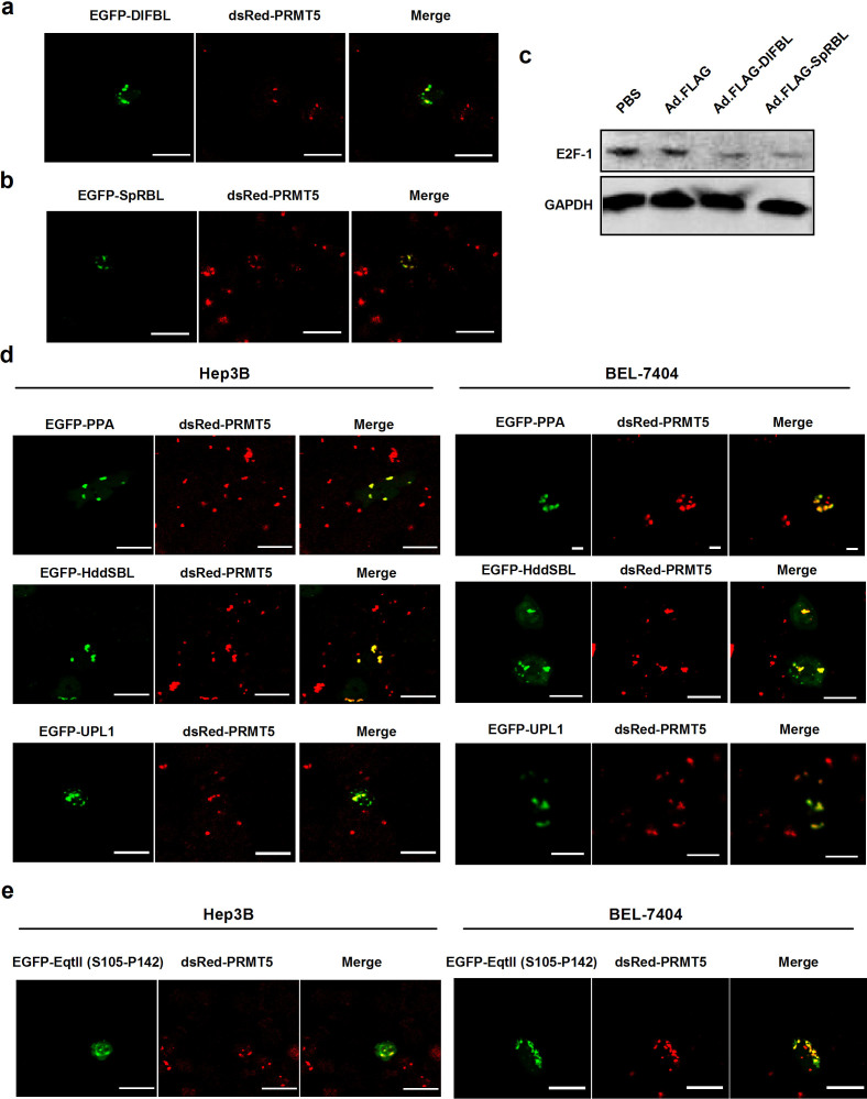
Figure 4. PRMT5 acts as a common target for various exogenous proteins.
(a) Hep3B cells were cotransfected with pEGFP-DlFBL-C1 and pdsRed-PRMT5. (b) Hep3B cells were cotransfected with pEGFP-SpRBL-C1 and pdsRed-PRMT5. (c) Hep3B cells treated with Ad.FLAG, Ad.FLAG-DlFBL, or Ad.FLAG-SpRBL at 20 MOI as well as PBS control for 48 h. Cell lysates were analyzed by Western blot for the levels of E2F-1. GAPDH served as the loading control. Full length blots were shown in Fig. S1. Plasmid pdsRed-PRMT5 was cotransfected into Hep3B cells or BEL-7404 cells with (d) pEGFP-PPA-C1, pEGFP-HddSBL-C1, pEGFP-UPL1-C1, and (e) pEGFP-EqtII (S105-P142)-C1. After 48 h, the co-localization of PPA, HddSBL, UPL1, and EqtII (S105-P142) fragment with PRMT5 was observed under a confocal laser scanning microscope. Bars show 20 μm.

