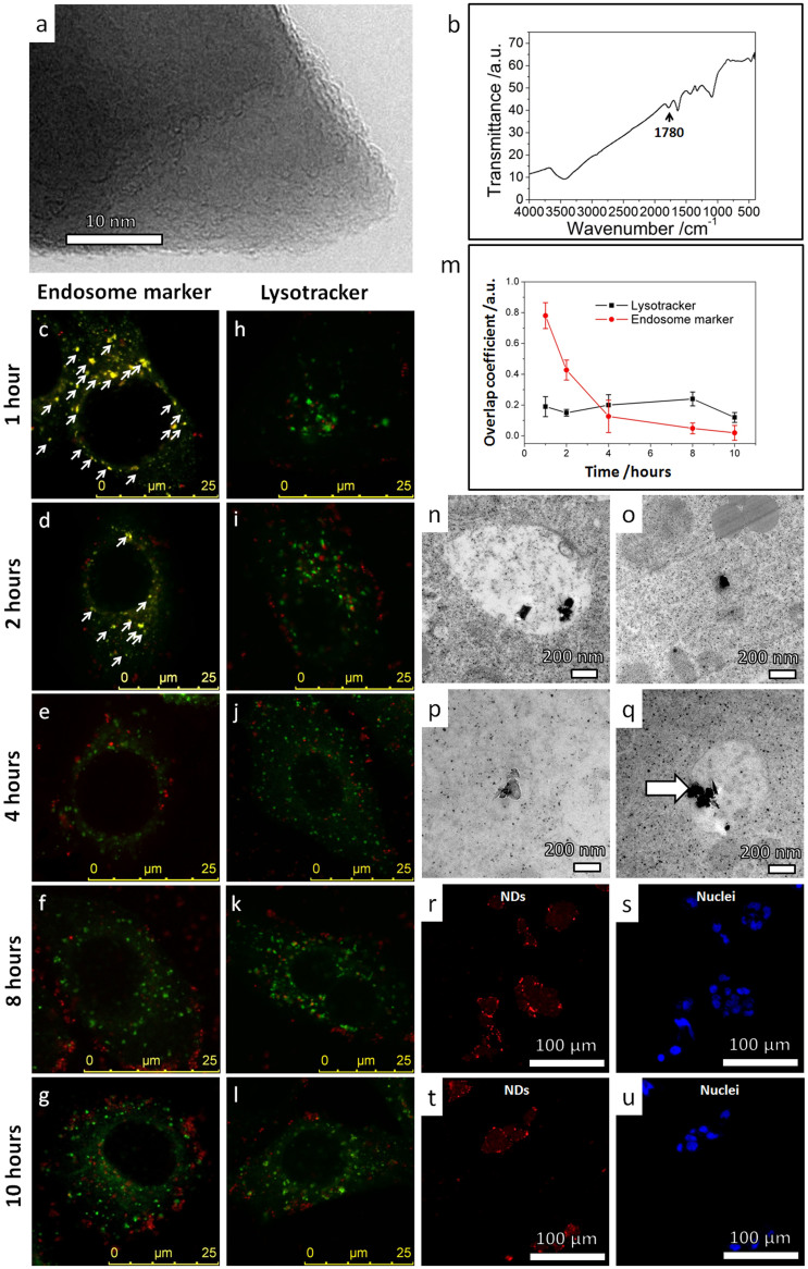
Figure 2. Intracellular translocation of sharp-shaped nanodiamonds from membrane bounded vesicles and stable residence in cytoplasm.
(a) High resolution TEM image showing the sharp corner of a typical nanodiamond. (b) FTIR spectrum of the nanodiamonds showing a characteristic dip (1780 cm−1) of a −COOH rich termination surface. (c–g) Representative confocal microscopy images showing the fluorescent signals of endosome markers (green) and nanodiamonds (red) in HepG2 cells after cell incubation with nanodiamonds in serum-free medium for (c) 1 hour, (d) 2 hours, (e) 4 hours, (f) 8 hours and (g) 10 hours. The yellow spots show spatial overlap between nanodiamonds and endosomes (as marked by white arrows). The observed low overlap for longer incubation durations indicates that the nanodiamonds escaped the endosomes. (h–l) Representative confocal microscopy images showing the fluorescent signals of lysotracker (green) and nanodiamonds (red) in HepG2 cells after cell incubation with nanodiamonds in serum-free medium for (h) 1 hour, (i) 2 hours, (j) 4 hours, (k) 8 hours and (l) 10 hours. Very low spatial overlap between nanodiamonds and lysosomes was observed, indicating that most nanodiamonds escaped the endosomes before the lysosomes were formed. (m) The overlap coefficients of nanodiamonds with endosomes (red symbols) or lysosomes (black symbols) functions of the incubation duration. (n–q) TEM images showing the typical intracellular distribution of nanodiamonds in HepG2 cells after incubation in serum-free medium for 24 hours: (n) TEM image of nanodiamonds residing in a membrane-bounded vesicle; (o, p) TEM images of nanodiamonds in the cytoplasm (majority); (q) TEM image of nanodiamonds piercing the membrane of a vesicle. (r–u) Representative confocal microscopy images showing the uptake and excretion of nanodiamonds in HepG2 cells as revealed by the fluorescence of nanodiamonds (red). The nuclei were stained by DAPI (blue). (r,s) HepG2 cells were incubated with nanodiamonds in serum-free medium for 12 hours. Punctated red dots show effective cellular uptake of nanodiamonds. (t,u) HepG2 cells were incubated for additional 12 hours in serum-free medium without nanodiamonds. The punctated-red-dot pattern remains unchanged, suggesting that most nanodiamonds remained in cells without being “washed out”.

