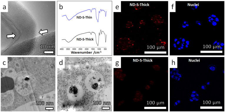
Figure 4. Intracellular translocation and cellular excretion behaviors of ND-S-Thick particles (nanodiamonds coated with thick layers of SiO2).
The observed cellular dynamics was similar to that of SiO2 nanoparticles (Supplementary Fig. S12 and Fig. S13). (a) High-resolution TEM image showing a ~15 nm thick silica shell on the surface of ND-S-Thick particles. The sharp corner of the nanodiamonds was rounded up by silica coating. (b) Similarity between FTIR spectra of ND-S-Thick and ND-S-Thin particles indicates that the two kinds of nanoparticles have similar surface chemistry. (c–d) Typical TEM images showing distribution of ND-S-Thick particles in membrane bounded vesicles, in HepG2 cells after incubation in serum-free medium for 24 hours. (e–h) Representative confocal microscopy images showing the uptake and excretion of ND-S-Thick particles in HepG2 cells as revealed by the fluorescence of NDs (red). The nuclei were stained by DAPI (blue). (e,f) HepG2 cells were incubated with ND-S-Thick particles in serum-free medium for 12 hours. Punctated red dots (fluorescent signal from ND-S-Thick) shows effective cellular uptake. (g,h) HepG2 cells were incubated for additional 12 hours in particles-free serum-free medium. Greatly reduced fluorescent signal from the ND-S-Thick (red dots) shows >75% ND-S-Thick particles exited the cells after the “washing” process.

