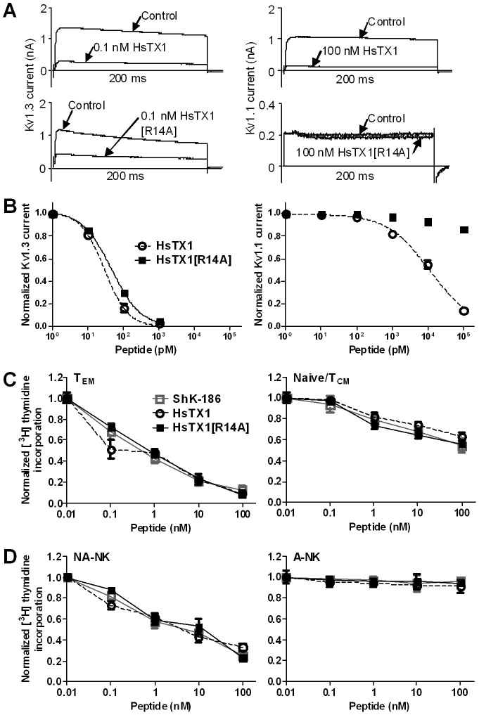
Figure 6. Functional analyses of HsTX1[R14A].
(A) Whole-cell Kv1.3 (left) and Kv1.1 (right) currents measured by patch-clamp in stably transfected L929 fibroblasts before (control) and after perfusion of HsTx-1 (top panels) or of HsTX1[R14A] (bottom panels). (B) Dose-response inhibition of Kv1.3 (left) and Kv1.1 (right) currents by HsTX1 ( and dashed line) and HsTX1[R14A] (
 ) fitted to a Hill equation (N = 3 cells per concentration). (C) Effects of ShK-186 (
 and grey line), HsTX1 (
 and dashed line), and HsTX1[R14A] (
 ) on the proliferation of rat Ova-GFP TEM lymphocytes (left) and of rat splenic T lymphocytes [mainly naïve/TCM cells] (right) measured ex vivo by the incorporation of [3H] thymidine in the DNA of dividing cells (N = 3). (D) Effects of ShK-186 (
 and grey line), HsTX1 (
 and dashed line), and HsTX1[R14A] (
 ) on the proliferation of human NA-NK (left) and A-NK (right) lymphocytes (N = 3).

