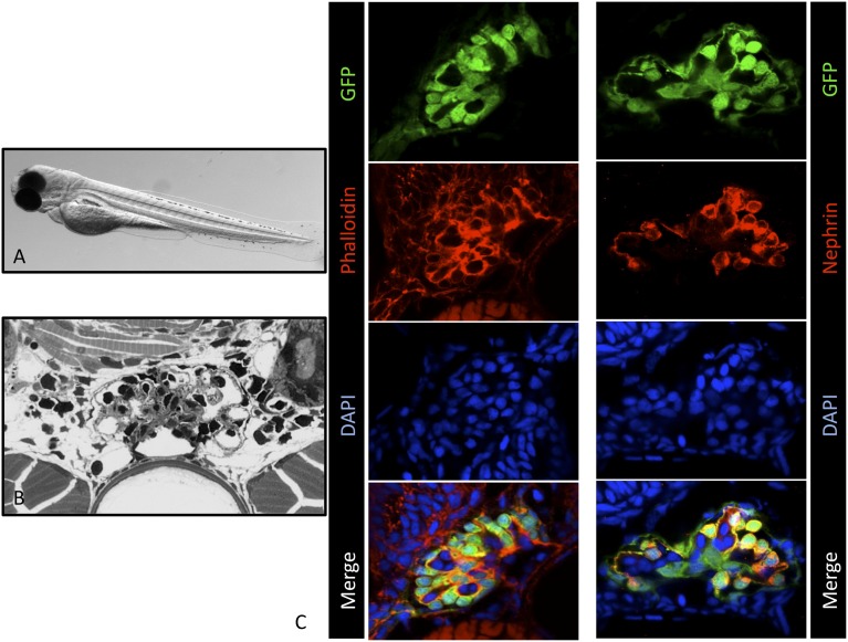
Figure 1.
ET larvae show regular glomerular morphology and nephrin expression. (A) ET larva at 3 dpf. (B) Semi-thin section of ET larva at 7 dpf. (C) Cryosections of ET zebrafish larvae at 3 dpf. Staining of F-actin by Alexa Fluor 546-conjugated phalloidin is shown in the left panel. The right panel shows the colocalization (yellow) of eGFP-positive podocytes (green) and the nephrin staining (red). Scale bars: 10 µm. DAPI, 4′,6-diamidino-2-phenylindole.

